A biopsy is a sample of tissue taken from the body and inspected under a microscope. A doctor performs a breast biopsy to remove tissue from a problematic location so that a pathologist may assess whether the sample contains malignant cells.
Previously, biopsies were only performed by making an incision in the breast and removing the suspicious tissue as well as some normal tissue from around it. The scars from these surgical biopsies may affect the size and form of the breast.
Doctors can now frequently employ novel procedures. Fine needle aspiration and core needle biopsy are two options that do not leave scars or alter the form of the breast. This is a substantial benefit because four out of every five women who have biopsies are cancer-free.
If your health care facility does not perform needle biopsies, seek to be referred to one that does, unless this operation is contraindicated for you.
What Is It Used For?
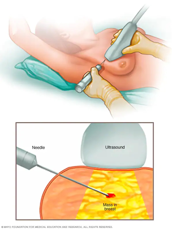
A bigger needle is utilised for a large core needle biopsy than for fine needle aspiration. Because the needle is bigger, more tissue may be extracted and inspected. Large core needle biopsy is frequently conducted using x-rays or ultrasound to ensure that the tip of the needle has reached the questionable area. It can aid in the evaluation of abnormalities revealed on a mammogram but not felt by touch.
If you have, a core needle biopsy may not be appropriate.
an abnormality at the chest wall, nipple, or breast surface particular types of calcium deposits in the area of concern very tiny breasts.
It can be difficult to obtain appropriate results from a core needle biopsy under certain circumstances. Instead, your doctor may advise you to undergo a surgical biopsy.
Preparation
To avoid bleeding issues, if you take a blood thinner, aspirin, or a nonsteroidal anti-inflammatory medicine (NSAID), you may need to stop using it for several days before the test. Tell your doctor if you are allergic to lidocaine or any local anaesthetic before undergoing the test.
How to Do It
You dress in a hospital gown that is exposed in the front. The core biopsy needle is about the thickness of a pen tip. It is frequently put into the breast via a small incision. The doctor guides the needle into the region of concern with x-rays or ultrasound imaging, or by feeling the lump. He or she uses suction from a syringe to retrieve one or more tissue samples through the needle. (Three to six samples are frequently retrieved for examination by a single puncture.)
If mammography or ultrasound will be used to guide biopsy needle insertion, all necessary equipment should be kept in the same room. There should be pressure but no pain. This operation only takes a few minutes.
Follow-Up
Following the core needle biopsy, your doctor may apply an ice pack to the incision for 15 to 30 minutes. You'll most likely resume normal activity practically quickly.
The biopsy sample needs to be examined, which typically takes several days. 65 per cent of women are given a benign diagnosis at facilities where biopsies are performed, allowing them to resume their yearly mammograms. Another 25 per cent are now receiving treatment for cancer or a precancerous disease. The following step is typically a surgical biopsy for the remaining 10% of cases where the results are inconclusive.
Risks
You can experience a little blood or bruise after the biopsy, along with some breast pain. For a scar, this surgery simply leaves a very small dot.
When to visit the doctor
People often simply need to call their doctors for the results of their biopsy because side effects are not expected.
Breastcancer.org
https://www.breastcancer.org
https://www.breastcancer.org
